Palmer: ‘PET Scan Not Appropriate As Initial Screening Tool’

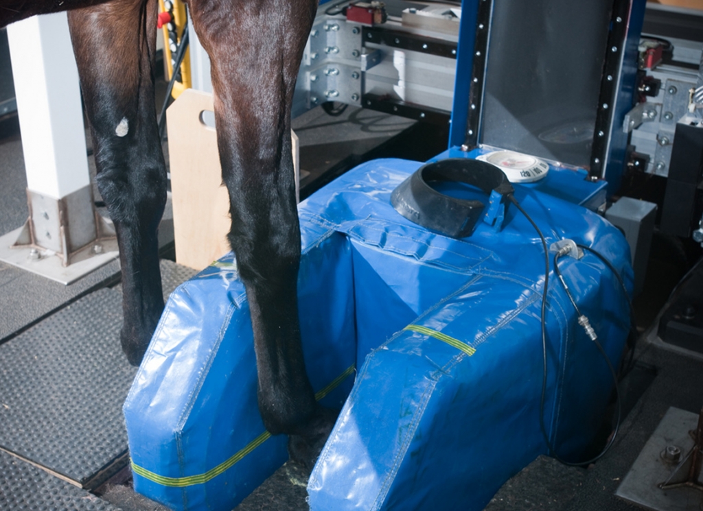
New York State Equine Medical Director Dr. Scott Palmer has described Positron Emission Tomography (PET) scan as a “fantastic diagnostic tool” and that “it can play a very important role” in helping to identify and pinpoint subtle musculoskeletal injuries in horses, but that the scan is not the best initial screening tool in singling out horses at risk for catastrophic injuries.

Palmer addressed the issue during an equine health and safety briefing held at Tuesday's meeting of the New York State Gaming Commission and also offered some preliminary findings  on the 17 equine fatalities recorded during this summer's Saratoga meeting.

“Identification of horses at risk for catastrophic injury must begin with a screening protocol that can be scalable, practical, affordable, and can be used by every horse while training or racing without regard to any clinical indication of injury,” Palmer said in revised comments issued Wednesday. “Wearable biometric sensors are best suited to accomplish this first level of screening. These sensors detect subtle abnormalities in a horse's gait that are not detectable with the human eye. They serve as a 'check engine' light that alerts us to the possibility that there is something wrong with the horse and that the horse should be examined by a veterinarian.

“The veterinary examination is the second level of screening for an abnormality that might predispose a horse to injury. The goal is to reach a diagnosis of musculoskeletal abnormalities and typically will include use of diagnostic nerve blocks and digital radiography. If lameness is detected in a limb during this examination and digital radiographs are inconclusive, then advanced imaging such as PET can be employed as a final screening step in this process.

“In summary, PET can play a very important role in the diagnosis of subtle musculoskeletal injuries in horses, but it is not useful at the initial screening level,” he concluded.

Palmer added that biometric sensors placed in horses' saddlecloths that can help identify at-risk animals, are “not quite ready for 'prime time' use” at this time.

In referencing the 17 Saratoga fatalities–a number that is approximately 1% of the 2000 horses stabled at the track but three times higher than 2021 and 2022–Palmer noted that the incidents were clustered around Whitney and Travers weekends. He added that fetlock injuries, typically responsible for 48-50% of fatal musculoskeletal injuries in New York and California over the last decade, represented 92% of all the exercise-associated fatal musculoskeletal injuries at Saratoga, a “significant finding” in Palmer's estimation.

Twelve of the 13 exercise-associated injuries occurred either during the final furlongs of the race or during the gallop out, suggesting that fatigue was also a contributing factor.

Palmer explained that the unprecedented 11 inches of rain that fell during the meet–compared to nine inches in 2021 and 8 inches in 2022–had a material impact on the consistency of the racing surfaces. During the meet, there were 65 surface changes (16%) compared to just 17 (4%) in 2022. With those facts in mind, “increased moisture in the Saratoga main dirt track and spatial and temporal variation of the moisture content of the track during the meet were likely contributing factors to the increase in the number of racing fatalities.”

A comprehensive report of the investigation will be made available to the public as soon as the investigation is complete.

The post Palmer: ‘PET Scan Not Appropriate As Initial Screening Tool’ appeared first on TDN | Thoroughbred Daily News | Horse Racing News, Results and Video | Thoroughbred Breeding and Auctions.

Source of original post

Screen, Scan, Save: Is This Racing’s Big Fix?

Like the wildfires fanned by this summer's hot winds, doomsday predictions of horse racing's demise have raged through the mainstream and trade press this year, fueled by a sickening spate of high-profile equine fatalities on the sport's highest-profile stages–tracks armed with some of the most stringent safety guardrails.

This means these horses passed before the eyes of a slew of experts–from the riders to the trainers to the veterinarians and the regulators–deemed among the best in the business. If they can't single out these horses before catastrophe happens, who can?

As the science around racehorse injury has evolved, the notion of a random “bad step” as the cause of catastrophic breakdowns has been largely debunked. Much cited since, a California Horse Racing Board (CHRB) led study from between 2011 and 2013 found that roughly 90% of Thoroughbreds that suffered catastrophic musculoskeletal injuries had pre-existing bone lesions near or at the site of the fracture.

The problem is, these lesions–abnormalities of the bone as it undergoes remodelling leaving that part of the skeleton vulnerable to fracture–can be a clinical nightmare to detect through the usual means. Think X-rays, nuclear scintigraphy, or just jogging the horse up and down a flat path. So, what to do?

Catastrophic injuries don't just happen overnight. The pathology underlying these events can occur over weeks, if not months, leading up to the fracture. Perversely, this is good news. There's a window for intervention, opening the door to a new breed of screening tools.

Much focus these past couple of years has been placed on a biometric sensor called StrideSAFE, which appears able to identify that small percentage of at-risk horses who are sound to the human eye. The New York State Gaming Commission equine medical director has called it “probably one of the most important contributions to the Thoroughbred horse industry that has ever been made.”

What's more, a new wave of imaging technologies are proving capable of diagnosing the underlying cause of these problems much earlier than ever before. Some of the industry's brightest minds say the trick to substantially reducing equine fatalities will be to standardize the combined use of these screening and diagnostic tools, bringing much needed objectivity to what–in identifying lameness–is all too often an exercise in subjectivity.

Easier said than done.

Logistical hurdles, significant costs and legitimate concerns about exactly how all this new information will be used present no small set of obstacles. But the imperative is clear, warn various industry leaders: If the sport doesn't act on these tools that it has at its disposal–and act quickly–racing could all too soon become an anachronism.

“This is the new frontier of racehorse safety,” said Jeff Blea, CHRB equine medical director. As an example, he highlighted Sleip, a smartphone app that has potential to work as a lameness diagnostic tool.

“StrideSAFE is the best screening tool we've ever had–clearly it's a lot more effective than the human eye,” said Warwick Bayly, professor of equine medicine in the department of Veterinary Clinical Sciences at Washington State University.

When it comes to integrating such screening tools into the daily furniture of the industry, Bayly was adamant. “We've got to do this.”
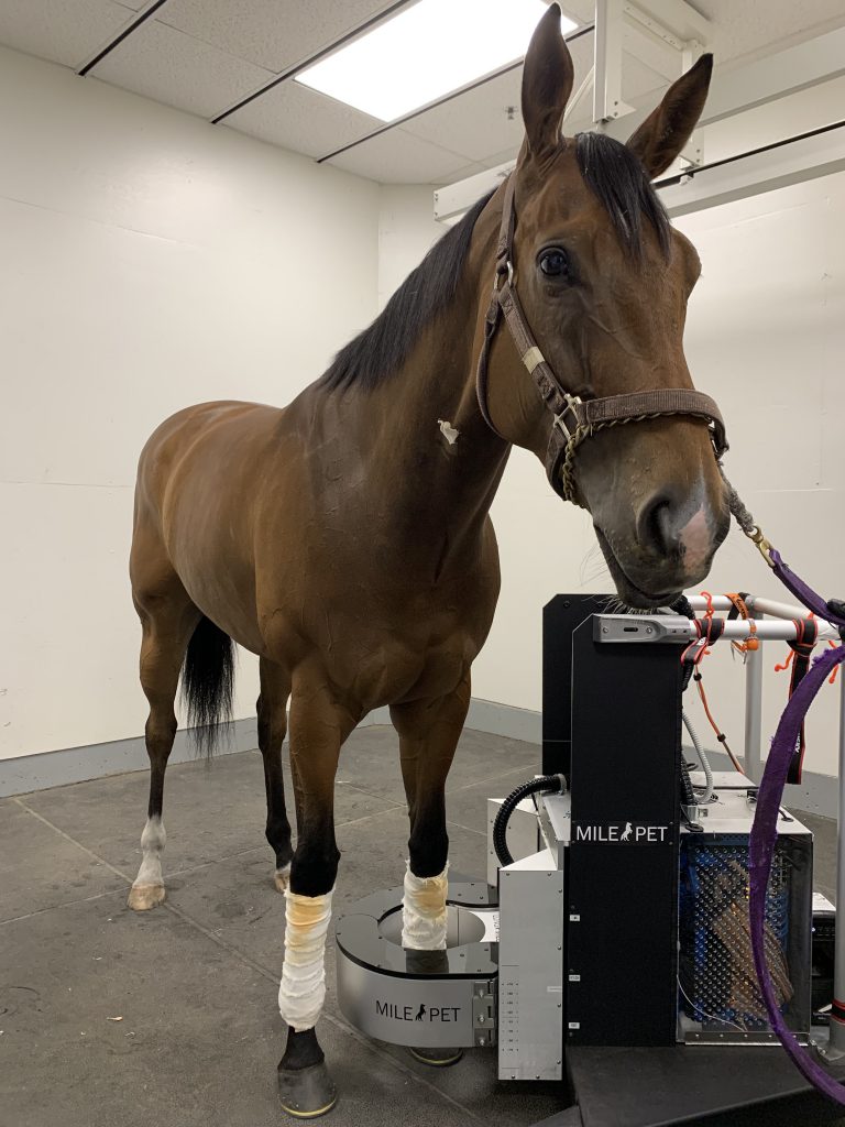
Positron Emission Tomography (MILE-PET) unit | courtesy Mathieu Spriet


“We cannot scan all horses to identify those four percent”

“It's got to the point where now it's more than a diagnostic tool,” said Mathieu Spriet, of the Positron Emission Tomography (MILE-PET) unit. “It is helpful as a clearance to race. It can help the regulatory vet by not scratching unnecessary horses. And it can help individual horses.”

The portable PET unit is the brainchild of Spriet, and has been a part of the diagnostic scene at Santa Anita since the end of 2019. Since then, nine other PET units have been distributed to Florida, Kentucky and Pennsylvania. In about one month, the University of Melbourne is scheduled to join the list.

In its relatively short life-span, evidence strongly suggests that the PET unit is especially useful in diagnosing the sorts of fetlock injuries–those actively brewing in the sesamoid bones, and the distal region of the cannon bone immediately above the fetlock–earlier and with greater accuracy than has been the case with more established imaging technologies.

Why is this important? The fetlock has long been the Thoroughbred racehorse's Achilles heel. Fetlock failures constituted in California nearly 60% of all musculoskeletal injuries that proved fatal during the 2018-2019 fiscal year.

In this study, a team of experts examined the results of 33 horses imaged using both PET and nuclear scintigraphy. The researchers agreed on the results for the PET more frequently than for nuclear scintigraphy. Indeed, they detected potential sesamoid bone problems in 22.2% of limbs imaged with PET, but only 6.9% of limbs imaged with scintigraphy.

In another study of 25 racehorses, 88% of the PET scans performed six weeks apart identified the same problem areas in the fetlock, demonstrating the technology's reliability. In all, 65% of the fetlocks examined demonstrated improvement during a 12-week rest period from racing.

But when it comes to the bête noire of horse racing–those at-risk runners who show no clinical signs of lameness–the study with perhaps the most significance concerns the 72 “normal” horses chosen to undergo PET scans of all four fetlocks at Golden Gate Fields, Santa Anita and Fair Hill.

Three of the 72 horses were laid up as a result of what showed up on the scans, while about 20% of the horses examined saw their training schedules modified to manage small underlying issues.

“We had one horse that potentially needed surgery,” said Spriet, of one of the three ostensibly “normal” horses found to have major underlying issues. “We know that there are some horses out there, clinically they are fine, but unfortunately they do break down.”

As Ryan Carpenter, a private SoCal-based veterinarian and a habitual user of PET, puts it, “I don't think there's any modality that's superior to PET when it comes to identifying problems in the sesamoid bones early.”

But what about other vulnerable parts of the racehorse's skeleton?

The same year Santa Anita welcomed the bespoke PET unit, it opened its doors to a standing magnetic resonance imaging (MRI) unit–a large, enclosed cell with dimensions of 10′ x 10′ x 30′.

MRI units, explained Carpenter, are especially adept at identifying issues in the lower ends (the condyles) of the cannon bone, including palmer osteochondral disease (POD), an all-too common degenerative bone problem in racehorses. “There's nothing better for POD than MRI,” he said.

For other notoriously vulnerable regions of the racehorse skeleton–like the shoulder region and the pelvis–Carpenter said that nuclear scintigraphy remains the go-to imaging modality.

There are drawbacks to MRI and PET. One is cost. A new PET unit will set you back some $600,000. Standing MRIs are typically leased from the manufacturer. According to Carpenter, the cost of scanning both front ankles with either PET or MRI is about $1,200.

Another is logistical. It takes between 45 minutes to an hour to scan both fetlocks with MRI.

“The knock on PET is that it requires a horse to go into an isolation period while it eliminates the radioactive isotope,” said Carpenter. The isolation time is about eight hours. “Here, they usually go over to the hospital for a PET at about five o'clock in the morning and they usually go back to the barn around two in the afternoon.”

Which leads to another imaging tool, computerized tomography (CT). Indeed, Racing Victoria requires all international horses that race there, including Melbourne Cup nominees, to undergo CT scans.

“The biggest benefit of the CT is what? It's fast. You can CT an ankle in 17 seconds. And there is no holding period,” said Carpenter.

But CT also has its drawbacks. It has yet to prove itself accurate at pinpointing the more worrying active bone remodeling as early as PET and MRI, said Carpenter. That said, CT offers promise for a diagnostic learning curve, he added.

“I'm hopeful that with CT we can eventually pick up the change in pixels in the location of the bone change before fracturing,” said Carpenter. “If you can make that true, you can scan all four fetlocks, use radiomics to look for changes in pixel density, and do what we're doing now with PET but in a fraction of the time.”

Given his experiences over the past few years, is Carpenter surprised by the results of the PET study that found 4% of 72 clinically sound horses had injuries that required them to be rested for an extended period?

“I'm not surprised at all,” said Carpenter. “We know those horses exist. Our hardest struggle as clinicians is determining which horses they are.”

Given the costs and logistical difficulties associated with PET and MRI, however, “we cannot scan all horses to identify those four percent,” admitted Spriet. “It's just not practical.”

But what if there was another way to objectively sift through the population to identify that small percentage of at-risk horses? Dave Lambert, founder of StrideSAFE, believes he's got the answer.
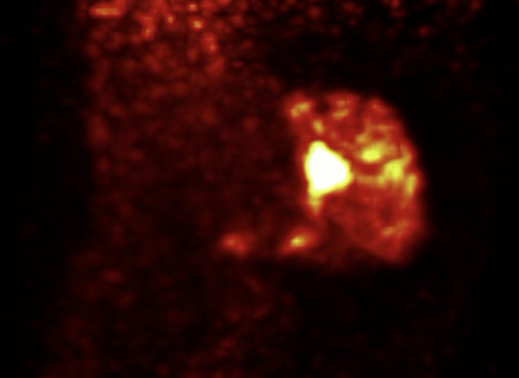
This scan image is from a fetlock with a severe sesamoid injury. 3D MIP is “maximal intensity projection,” which is a true 3D rendering.


“This is exactly how the system was designed to work”

StrideSAFE is a discreet bio-metric sensor used on horses working or racing to capture a variety of measurements related to the horse's acceleration and deceleration, its up and down concussive movement, and its medial-lateral motion. In other words, the horse's movement from side to side.

In short, StrideSAFE has proven capable of detecting the sorts of subtle abnormalities in gait even the most seasoned trainers, veterinarians and exercise riders miss when watching horses jog up and down a path, or when taking them through their paces.

StrideSAFE originally worked on a traffic light system, with a green for all-clear, a yellow for caution, and a red for possible danger.

In a long-term study conducted on horses racing at New York Racing Association (NYRA) tracks, of the 20 horses that suffered fatal musculoskeletal injuries during the period of the trial, 18 of them had received a red rating in a race before suffering a catastrophic breakdown, said Lambert. One of the 20 had received a prior dark amber rating.

These red and dark amber ratings were issued in either the race immediately prior to the breakdown or else two or three races back, said Lambert, meaning StrideSAFE detected 90% of those horses that suffered a catastrophic injury sometimes weeks or even months in advance.

In total, about 15% of the horses involved in the study were red-flagged, said Lambert. Given how at that time there was still much to learn about StrideSAFE's efficacy, there was no coordinated system to funnel those red-flagged horses for follow-up diagnostics.

Since then, Lambert and his team have refined the system to what he calls a “risk factor calculation” from one to five. Five is the category in which a horse is most at risk of a fatal or career ending injury–nearly 300 times more likely than horses that fall within risk category one. In all, 73% of the horses fell within category one, the safest group.
David Lambert & trainer Dale Romans | StrideSAFE

Using data from the same 6,616 individual starts in the NYRA study, Lambert determined that about 5% of the horses studied–a number totaling 363 horses–fell within the risk category of five. That includes the same 20 horses that suffered catastrophic injuries, 18 of which were red-flagged in prior races.

If the other two fatally injured horses had worn StrideSAFE in high-speed workouts between races, would the sensor have picked up a problem? “We don't know for sure,” said Lambert. “But there's a good chance it would have.”

Interestingly, Lambert said that in general, the red-flagged horses raced back quicker than horses in safer categories. “These horses are sound and they're ready to race,” he said, “but they're sitting on a fracture.”

Earlier this year, StrideSAFE was used on horses racing at Churchill Downs's short Spring meet. All eight horses that suffered a fatal race-day musculoskeletal injury were carrying the technology.

Seven of the eight musculoskeletal cases showed abnormal sensor readings as soon as they left the starting gate, prompting Lambert to subsequently remark that “had the sensors been on the horses in prior races, they could have pointed to an issue the horse was having weeks or even months earlier.”

Unlike the NYRA study, at Churchill Downs there was a more coordinated system for following up with the trainers of flagged horses. StrideSAFE flagged two visibly sound horses that were subsequently sent for PET scans, revealing the beginnings of condylar fractures.

“This is exactly how the system was designed to work,” explained Lambert. “Screen every horse in a race, detect those at high risk, diagnose using modern technology and bring about a cure rather than suffer a fatality.”

But not every horse that's categorized as being at highest risk of injury harbors an underlying issue. And so, to refine the system even further–iron out the kinks–what would be the best application of StrideSAFE?

“If every horse in training wears it,” said Will Farmer, equine medical director for Churchill Downs.

“If every horse had this for every high-speed work and in every race, I think that would be the ultimate goal,” Farmer added. “Anytime that a horse reaches a high speed, and they have a sensor on their back, we would have such a complete picture, there would be multiple opportunities for veterinary intervention.”
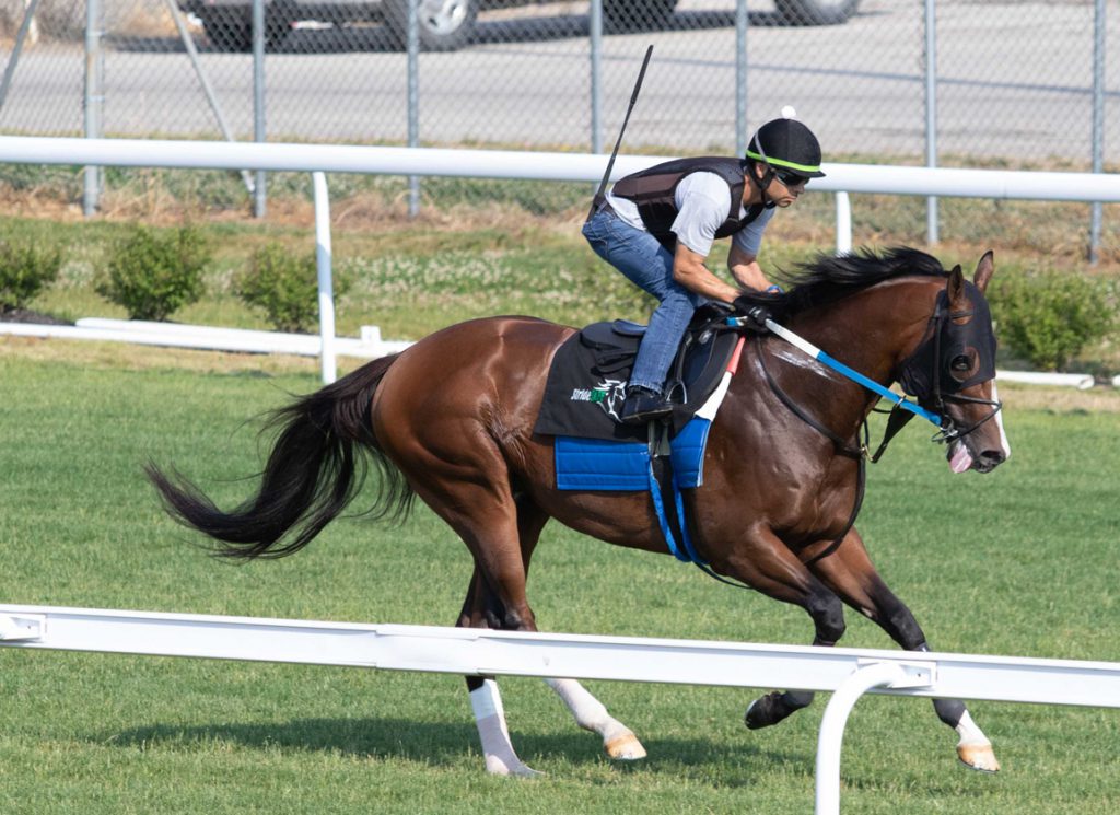
A Steve Asmussen-trained worker equipped with StrideSafe | Holly Smith

The results from StrideSAFE present an interesting parallel with a recent study out of Tasmania, which found that horses will decrease their stride length in the weeks leading up to an injury.

Back in 2010, Tasracing partnered with Stridemaster, which had developed a biometric sensor technology, to provide a race-day timing system using GPS and motion sensor data. The original purpose was to provide information to share with the betting public.

“We said, 'hey, that looks like great data. Can we get a hold of it?'” said Chris Whitton, professor of equine medicine and surgery and head of the Equine Centre at the University of Melbourne.

Whitton and his fellow researchers reviewed the data from 584 different horses who made 5660 individual starts. They found a “marked rate of decline” in speed and stride length roughly six races prior to injury.

Going into the study, “I would have thought it was one or two races that they would start showing effects,” said Whitton. “Such a long period I thought was fascinating.”

The researchers also found that horses are especially vulnerable to injury early in their racing career. “The first and second race of a horse's career is actually quite a high risk,” said Whitton.


The Heart of the Problem

Fractured bones don't constitute the only cause of lost equine life on the track.

Though fairly rare, sudden cardiac deaths occur at a rate of one for every 8,789 starts racing, and one death for every 158,000 Thoroughbred training days, according to this CHRB report. But what exactly causes these events largely remains a matter of debate.

This well-considered study looked at post-mortem findings from 268 Thoroughbred racehorses that suffered exercise-related sudden deaths. Pathologists made a definite diagnosis in only 53% of cases, a presumptive diagnosis in 25% cases, while 22% of cases were left unexplained.

Which leads to another biometric sensor technology finding its niche in the sport from a company called Arioneo.

“In total, we are working globally with about 600 trainers and vets,” said Valentin Rapin, Arioneo's managing director. “In the U.S. it's really the beginning for us. We're working with about 40 trainers at the moment.”

Like StrideSAFE, Arionoe's Equimetre fits onto the horse's tack during exercise, sending information back to the user, who can view it on a user-friendly computer dashboard. Part of that data concerns the horse's stride length and stride frequency at all gaits, from walk to trot to gallop and workout.

But Arioneo's Equimetre–which costs about $190 a month per device–also monitors heart-rate, providing data that can be used to assess both performance and overall health, including whether the horse is experiencing physical pain or discomfort, said Rapin.

“We can also look at the level of effort the horse is reaching during exercise. Sometimes you will see some abnormally high level of heart rate during exercise, depending on the speed the horse is going. And you can also see some abnormal recovery data,” said Rapin.

If the heart-rate monitor sends out what amounts to red flags about the horse's heart health, an ECG electrocardiogram (ECG) monitor can be used to map a more sophisticated picture of the horse's heart rate and heart rhythm, to identify any possible underlying problems like arrhythmias.

Rapin highlighted a horse in Australia routinely equipped with the monitor until one day in a workout, its heart rate spiked unusually high. A subsequent ECG analysis of the horse prompted its retirement.

Cristobal Navas De Solis, assistant professor of cardiology/ultrasound and internal medicine at the University of Pennsylvania School of Veterinary Medicine, is one of the researchers involved in an ongoing study using Equimetre on three-day-eventers during exercise.

The goal is to find out “which variables are more associated with injury or performance,” said Navas De Solis.

From a recent preliminary analysis of the first two years of data–involving 53 horses and 1,467 individual workouts–the researchers have identified several variables that appear to have some relevance on performance and injury: Heart rate and stride rate during the warm-up period, stride length at certain speeds, symmetry at trot, and heart rate recovery after a workout.

Navas De Solis sees early markers of injury as the “sport's holy grail,” and the standardization of screening tools like Arioneo and StrideSAFE in horse racing as “a big step forward in this direction.” He highlighted an Italian pre-screening program for human athletes that decreased sudden deaths by nearly 90% after implementation.

But he's also a pragmatist. To get to that stage, racing's leaders would need to agree upon a set of well-designed but probably expensive studies involving thousands of horses.

The ultimate program would be one that weeds out the most at-risk horses, and similarly ensures that perfectly healthy horses aren't excluded from competing via false positives.

“Having an arrhythmia during exercise doesn't necessarily mean that the horse or the athlete has a problem. From an arrhythmia standpoint, it's not an all or nothing. Some arrhythmias are okay during exercise and are not going to cause a problem to a horse,” Navas De Solis said.

A key question therefore, said Navas De Solis, is “how do we find the right balance between decreasing injury and not causing unjustified alarm?”
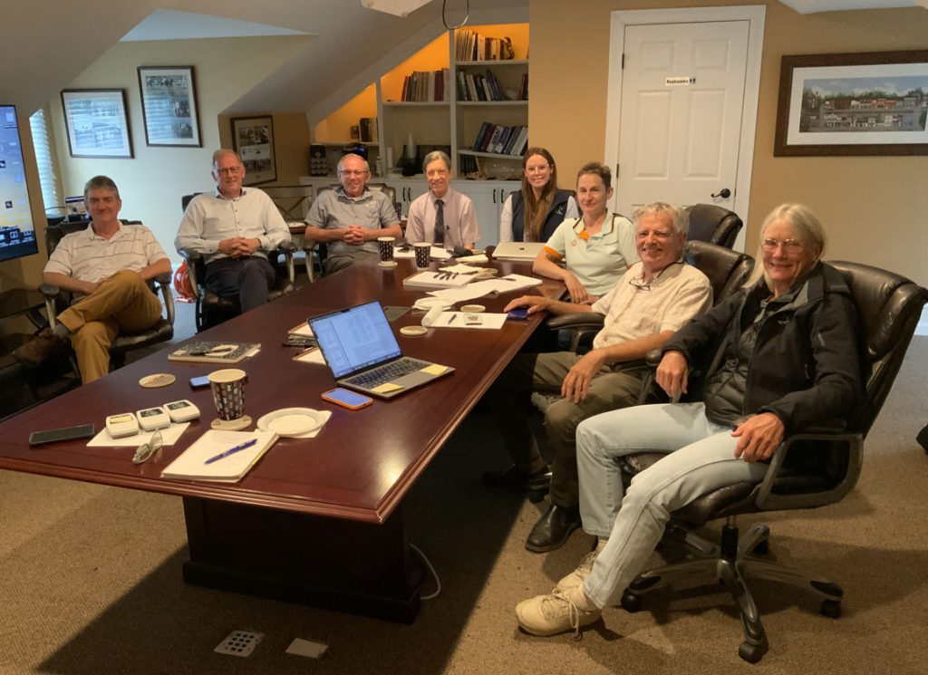
The Research Team at StrideSafe | StrideSafe


“Nothing that can't be solved”

For Farmer's vision of StrideSAFE being used on every horse that works to materialize, a key obstacle is one of logistics.

“Race days are very structured,” said Farmer, of its main use to date at Churchill Downs. “We know times. We know distance. We know horse names. We know everything we need to know days in advance.”

The organized chaos of a busy training morning–with hundreds of horses working, many simultaneously, from dozens of different barns–makes the practicalities of using StrideSAFE infinitely harder in comparison, said Farmer.

“Right now, the logistics of training are very challenging,” said Farmer. “When you collect that data, you then need to identify which horse that refers to. In the sensor design as of today, it's not geared for you to just put it on their back and go. But it's nothing that can't be solved.”

Lambert acknowledged these logistical hurdles but called it “simply a matter of money” to get fixed–at the same time, noting significant contributions already to StrideSAFE from Jim “Mattress Mack” McIngvale.

“The technology is all there, we even have the idea envisioned, we have planned it out in readiness,” Lambert said, adding that incorporating AI technologies to the mix could help reduce the current costs of $35 a day per horse.

These screening tools also raise other questions to grapple with. For one, who should have access to the data generated? Another is cultural–a sense among those in the industry forged on tradition and custom that these devices could unnecessarily handcuff a trainer.

“Regulations are not going to eliminate risk,” Eric Hamelback, CEO of the National Horsemen's Benevolent and Protective Association (HBPA), at a town hall about StrideSAFE earlier this year. “And quite frankly, we don't want regulations to eliminate horsemanship.”

Proponents of StrideSAFE and Arioneo stress the ability of these technologies to complement the art of training, not to replace it.

“It's a tool that provides objective data, and the goal at the end is to combine the expertise of the trainer–who has all the knowledge and the feeling and the expertise about the horse–with the data to make it successful,” Rapin said.

That idea taps into what Mathieu Spriet means when he talks about “voluntary longitudinal follow up” with imaging devices like PET to make doubly-sure a horse is up to the rigors of training and racing. In other words, the more horses are scanned periodically, “I think the better that is for managing the career of a specific horse,” said Spriet.

“Connections can be like, 'well, this horse is doing well. But we have a big campaign ahead of him, so let's scan and see how it goes,'” Spriet added. “To me, this is the most exciting part of it because we can see things [you ordinarily couldn't] to better manage horses throughout their careers.”

Trainer Dan Blacker is representative of the new generation of conditioner who has embraced the imaging tools in Southern California. He said he's interested in the idea of using StrideSAFE on his horses. “For sure it's got a place,” he added. “If they bring it to California, I'm very willing to support it.”

Dan Blacker | Benoit

Interestingly, The Stronach Group is working on its own screening system which uses high-definition cameras to create detailed skeletal movement maps of horses.

But in the short-term, said Blacker, PET holds the potential to be a major “gamechanger” in the fight to reduce equine fatalities due to its accuracy and simplicity of understanding.

“The thing with the PET scan, it takes away the guess work. That's the one thing I want to get across to people in racing,” Blacker said. “It gives you a clear-cut answer to the question of whether you can keep racing or not. Once trainers nationally get to see that, you're going to see a lot of improvement.”

The post Screen, Scan, Save: Is This Racing’s Big Fix? appeared first on TDN | Thoroughbred Daily News | Horse Racing News, Results and Video | Thoroughbred Breeding and Auctions.

Source of original post

The Bare Bones: a Primer with Dr. Bramlage

He hasn't got all day; nor, doubtless, do you. So let's cut to the chase. We won't dwell on the journey that has made Dr. Larry Bramlage a doyen of orthopedic science, in its daily application to the racehorse: not the alphabet soup of honors and distinctions, nor the long experience that has honed the sharpest diagnostic eye in the business through 23 years with Rood and Riddle. We have simply dropped into the clinic, on a recent visit to Lexington, to direct a brief sunbeam of his knowledge and insight into the practices of those who depend for a living on the miraculous but fragile equilibrium of the bones that support a Thoroughbred.

“Racehorses are so good because they produce their skeleton based upon what they do,” Bramlage begins. “They're not born with it. Their skeleton is the minimum weight that they can produce and still carry them around the racetrack. So they have a big engine, but their undercarriage is no heavier than it needs to be. And that's why they're fast.”

That's true, in some evolutionary measure, of all horses–and other animals, too, people included.

“The skeleton is different than hearts and lungs and muscles,” Bramlage explains. “Those train to a volume of work that you're doing. Skeleton trains to the level of work that you do.”

He recalls a series of experiments conducted on turkeys some years ago, where one wing was restricted and the fowls learned to flap the other to get food. The idea was to establish how many cycles of this activity were required to stimulate bone.

“Well, it's interesting,” Bramlage says. “Because when you reach 36 cycles in a day, that's the maximum the bone will respond to. You can go to 2,000 and it won't get any stronger than in those 36. And that's what makes a trainer's job tough. Because they have to push the horses hard enough, that they get strong enough to carry themselves around the racetrack. But if you do too much, then those extra cycles begin to be destructive.”

Those 36 cycles, for our purposes, apparently equate to about a furlong. Which, Bramlage explains, means that your fastest eighth will be the level for which your horse produces bone. Obviously that doesn't happen overnight, albeit bone is far more dynamic than most laymen assume.

“But the stimulus is there that it'll try to reach that next level before the exercise does,” Bramlage explains. “And then you repeat that over and over, and eventually the skeleton gets appropriate enough that you don't acquire any damage during those 36 cycles. So while there's some always ongoing wear-and-tear, the most important part of making a racehorse is usually up to four or five races. Once they get there, their skeleton is virtually made.”

The living nature of bone, however, does mean that the “made” skeleton can regress once taken out of training. But Bramlage is keen to address a misapprehension, which took root maybe a decade ago, that persistently galloping a young horse creates the foundation for a strong skeleton.

“Galloping a horse a lot helps the heart and lungs–but once you go past those 36 cycles in a day, the rest of them are just wear-and-tear,” he explains. “A lot of horses were actually harmed by excess galloping.”

Previously there had also been the attempt to extrapolate the principles of interval training, in human athletes. “I knew a couple of people who, as runners themselves, were going to interval train and beat everybody,” he recalls. “And they ended up with 4-year-old maidens with splints on their hind legs. Because the skeleton just can't take that that many fast intervals. In people, the limiting system is the heart and lungs, not the skeleton. Horses have such great heart and lungs that, unless they're bleeders, they virtually never limit. The horse's heart and lungs can respond to anything you throw at them. But the skeleton has to do it in little stair steps. And that's how, in young horses especially, the heart and lungs often get ahead of the skeleton.”

Though the tibia also registers trouble here–it absorbs a lot of force, in locking the reciprocal motion of stifle and hock–the most familiar symptom is shin trouble.

“You go too fast, the wear-and-tear begins to exceed the response and you get bucked shins,” Bramlage continues. “Shins have to triple in size. The front cortex of a cannon bone in a 'made' racehorse is three times thicker than in the yearling that started training.”

So how does this translate, ideally, into building up a young horse towards a race? Bramlage suggests a pretty familiar scenario: one or two furlongs at a rather higher level than the rest of the exercise, in effect showing the skeleton where it's going to be asked to go in three days' time. The real skill, in training, is monitoring attitude.

“People ask, what makes a good trainer?” he says. “For me, it's an easy question. It's being able to understand when the horse is happy and when he's not. When horses are adapting well, they're happy to train. When a horse starts not wanting to go to the track in the morning, not wanting to load in the gate, those are the kind of things you need to look out for. It's a real art for trainers to understand when to push a horse and when to back off.”

Obviously you would hope that trial and error, over the generations, should have brought horsemen's intuitions pretty close to where they might land through learned science.

“If you go back to when Aiken, South Carolina, was the winter training center–because that's how far the railroad went south–they would have the Aiken trials and those 2-year-olds, early on, would be breezing an eighth,” Bramlage says. “Those short breezes were actually very useful to the horses. Especially when you're making the horse, it's a matter of trying to train heart and lungs–because you have to do that–without overtraining the skeleton.”

When a horse is past that stage, but has to be laid off training, the skeleton will not lose much strength through the first month but the situation will change pretty rapidly after two months. And a more significant spell, say four months, notoriously invites humeral or tibial stress fractures in a small number of horses: again, because heart and lungs train back so much faster than the skeleton.

Needless to say, by the time a horse is sent into the clinic, they have typically signaled a loss of form.

“If a horse has swelling in a knee or ankle, those guys at the racetrack pick it up,” Bramlage says. “These horses [sent into the clinic] don't have any obvious pain, heat or swelling, but their form has gone down. And a lot of times they have either bilateral lameness–two fronts or two hinds, sometimes all four–or they're just early wear-and-tear injuries. I think most of the really successful trainers today understand better than they did 10 years ago that the horse is subject to that wear-and-tear; and that whenever a horse is not giving you what it can, then you need to start looking.”

Parallel advances have been made in imaging technology. It is barely 30 years since radiographs were still processed on celluloid. Digital radiographs have themselves improved dramatically, and now scanning in three dimensions via CAT and MRI and ultimately PET is available.

“Nuclear imaging was a huge tool because those scans allowed us to look for stress fractures that didn't have any outward clinical signs,” Bramlage says. “But whenever regulators think in terms of needing a PET scan to monitor horses at the racetrack, that's not really true. You need to look at them and identify the horse that needs to be looked at, not scan them all. Most of those can be unraveled using all the tools we currently have. It's just a matter of knowing when you need to look. And so more than we need more equipment, we just need to look more often.”

The role of regulatory veterinarian is a contentious and evolving one. The process is being aided, however, by a growing injury database to succeed anecdotal assumption. Already Bramlage can see where this might take the profession.

“It may not hit during my lifetime, but I think the next really exciting revolution, which is going to totally change our care of racehorses, is digital timing,” he says. “It just makes sense that it will eventually move away from clockers and all be done passively, automatically, by the equipment. Well, when you have that data, it's not a real hard step to write an algorithm that identifies [problems that may be brewing].

“You could look at a horse's exercise fingerprint because stride length and stride cycle is pretty stable for individual horses. When the length begins to shorten, he's protecting something. And so each horse will have his own digital fingerprint, and this will be automatically recorded every time a horse works, every time they race. And all of a sudden you can say, 'This horse is getting into trouble.'”

Some early research has detected patterns that might anticipate injury as many as three races ahead. Bramlage can see a future where every horse will transmit data to central monitoring for red flags. For now, until the necessary technology is available, it falls to people like Bramlage to determine the level of risk that warrants its prohibitive cost.

“But I think that in the next generation beyond me, that will become automated,” he predicts. “And that will revolutionize the prevention of injuries. It'll be the best thing that ever happened.”

And that's one of the things that maintains such youthful enthusiasm in a septuagenarian who has already witnessed such transformation in the tools of his trade: the curve is only going to steepen.

Aside from digital radiography, the biggest leaps forward have been internal screws and plates; plus arthroscopy and its adaptation from diagnosis to treatment. For internal fixation, the initial debt was apparently to a Swiss cost-benefit analysis of chronic disagreement between tibias and ski-boots. Of arthroscopy, meanwhile, Bramlage muses: “Surgery never used to happen until there wasn't anything else you could do. Then with the arthroscope it became easier, quicker, better. And so now that is the first line of defense. The horse gets a chip fracture, they take it out right away. The joint doesn't degenerate, they go back to normal.”

Horsemen nowadays have gained faith that condylar fractures can be routinely secured. One of Bramlage's most celebrated patients, Personal Ensign, went a long way to changing perceptions. Nowadays you'll find many a Breeders' Cup winner with a screw lurking somewhere in its skeleton. It's a very different world from when Bramlage started out, and yet he feels we have barely started.

“Yeah, we were dipping X-rays in chemical solutions when I was a student,” he reflects. “But the young veterinarians right now will probably see the same explosion. Probably in the biologic areas: the understanding of cell biology, and cell communication, is doing the same ramp up. The ability to treat is going to be much more pointed and effective than now.”

Bramlage is acutely aware of our industry's exposure to an ever more urban society that professes ever fiercer vigilance on behalf of animals with which it typically has little interaction, certainly compared with generations past. In that respect, veterinary regulation manifestly has a front-line role. He's excited, then, that a digital fingerprint might give mute animals a new way of telling doctor what's wrong.

Even with the advent of such tools, however, Bramlage believes that the essential mystique of the Thoroughbred will endure. We might be able to explain how everything fits together, and learn how to put things back together, but the key to performance will remain elusive.

“And actually I hope we never do get to that point where we understand everything about a horse,” he admits. “Because I think that's what's intriguing to people. You can improve your odds by improving your breeding. You can keep the horse healthy, you can have a trainer that's capable to that level. You can do all those things, but you still can't just go buy a Derby winner.

“Every horse is a product of a dip out of the gene pool. It's not a one-to-one combination of the mare and the stallion. There are all sorts of units. Like you've got four genes that cause eye color in people. There are all those different combinations of things. So to combine whatever comes out of that gene pool with the mental capacity, to train hard enough and compete hard enough, you never know which horse is going to have it.”

He chuckles, and asks whether you ever heard of a racing mule named Black Ruby?

“Well, she was on the California fair circuit for about 10 years and there was only one other mule could occasionally beat her,” he explains. “But they cloned her several times, and none of them could beat me. They had the exact same genetic makeup, but none of them would run like that. So that elusive factor, I think, is what keeps people intrigued. And I hope we never identify that.”

Even his exceptionally intimate professional relationship with horses, ranging from Personal Ensign to claimers at Ellis Park, has only marginally clarified the enigma.

“I don't know that there's any one thing,” he says with a shrug. “Good horses are always physically attractive, well balanced. They're almost always smart, they're very intelligent, very adaptable.”

Does that make better horses better patients, too?

“Absolutely,” he replies. “But racehorses are the best patients anyway, in my opinion. The worst patient is the 4H horse that's never felt anything but a rub rag, because when they have to deal with pain, you never know how they're going to handle it. But racehorses are just like people who train hard: you're stiff and sore next day and then it goes away and you feel better than you did before you started. They have better survival instinct.”

And while recruitment to equine practice is becoming harder, given the reduced social exposure nowadays between young people and horses, Bramlage guarantees endless fascination to the next generation. The measure of your work, he says, is so much more gratifying than in small animal practice.

“I think equine practitioners tend to practice a lot longer because there's another level of assessment,” he says. “Your horses have to go back and run. They have to win barrel races. They have to win ribbons, if they're a backyard horse they have to trail-ride. There's a couple of books I read, discussing why do armies fight? It's mostly not for abstract ideals. They fight for the people next to them, the people they trained with, the things they know and the fear of failure. And I think this level of assessment, with the possibility that you'll fail, but the rewards when you succeed, it's higher in horses.”

And there are literally hundreds of horsemen in the Bluegrass who will be relieved to hear him say that. “I could easily be retired,” he says. “At some point, physically it's not going to be possible to continue. But I think that's why people stick around. I mean, when the success barometer is the dog being able to get up on the sofa? That's not quite as intriguing!”

The post The Bare Bones: a Primer with Dr. Bramlage appeared first on TDN | Thoroughbred Daily News | Horse Racing News, Results and Video | Thoroughbred Breeding and Auctions.

Source of original post

New SoCal MRI Study Aims to Bring Clarity to its Diagnostic Role

Seeking a full stop to the spate of high-profile fatalities in the race that stops Australia, Racing Victoria this year tightened the veterinary screws. The practical rollout of these efforts can hardly be described as an unadulterated success, however.

One of these new measures was a precautionary CT scan of all runners in the days leading up to the G1 Melbourne Cup–a target that hit the skids when Racing Victoria's new $1.27-million CT unit suffered a malfunction with the Cup field only half scanned, leaving the rest to be X-rayed (with a machine that was also temporarily incapacitated).

But other, less-mechanical incidents highlight some of the more nuanced problems that come with using sophisticated–and still yet relatively new–imaging technologies to diagnose lameness in equine athletes.

Despite the results of a mandatory CT scan that gave French import Gold Trip (Fr) (Outstrip {GB}) the all-clear to train up toward the G1 Cox Plate, Racing Victoria's veterinary team scratched him on the eve of the race–a diagnosis that jarred with the horse's connections, who declared him sound.

In a further twist, Gold Trip was given the all-clear to run in Sydney in the Rosehill Gold Cup just a week later–only to be scratched once more due to the prevailing firm going.

In short, as more and more regulatory veterinarians turn to imaging modalities like MRI, PET, and CT to help diagnose lameness, they're left to wrestle with slippery conundrums.

What clear connection exists between the image before them and an increased chance of injury in the horse, for example? And without an extensive historic medical record at their fingertips, how can they be sure that any possible abnormality that appears on the image is significant?

A new standing MRI-focused study set to launch in Southern California seeks to provide some much-needed answers.

“Lameness is a precursor to fetlock failure, and maybe we find bone changes that help us identify lameness. But we should never get to the point where the fetlock fails–we want to do better than that. And that's the goal of the study,” said Florida-based John Peloso, the lead researcher on the study.

“We need to figure out when they're helping us,” Peloso added, of imaging modalities like the standing MRI. “We need to learn more.”

Standing MRI | UC Davis photo

Participants in the study–which is funded by the Dolly Green Research Foundation and the Southern California Equine Foundation–will be split into two.

There will be 23 case horses whose lameness has been narrowed to the fetlock region, and 23 control horses who exhibit no visible sign of lameness.

The 23 case horses will be selected by Dr. Tim Grande, the California Horse Racing Board (CHRB)'s chief official veterinarian, from a variety of scenarios where regulatory veterinarians commonly have to intercede in a horse's training or racing program.

These include a morning or race-day scratch, a voided claim, and lameness in the test barn or following a scheduled work or race.

The control horses–those with no visible lameness–will be selected as a comparative match in terms of things like sex, age, and class.

If a case horse is picked from a race, then the winner–if sound–will make an obvious control match. If a case horse is selected after a workout, then a suitable match will be selected using PPs.

And what exactly will the researchers be looking for? The answer encompasses four specific areas of concern within the fetlock joint, the primary site of musculoskeletal injury in racehorses.

Researchers will be looking for density within the proximal sesamoid bones and distal cannon bone, bone marrow edema–or swelling–in the cannon bone, and palmer osteochondral disease, a type of bone bruising commonly referred to as just “POD.”

To elaborate on these points, Peloso pointed to a couple of relatively recent papers he had co-authored connecting important diagnostic dots.

Two issues associated with fetlock failure are high density–noticeable bone development that predisposes a horse to a greater risk of fracture–in the sesamoid bone and palmer osteochondral disease, while condylar fractures are linked to bone marrow edema and high density in the distal cannon bone.

“It's because of those two papers that we've dialed in on those bone changes,” said Peloso. “Maybe the study will teach us something new, and so, there'll be something that gets added to it.”

The standing MRI unit has been part of the Southern California backstretch furniture since the start of last year. Since then, the unit has been used to scan hundreds of fetlocks.

Nevertheless, as a relatively newfangled diagnostic tool, the MRI is still looked upon with a touch of skepticism by some corners of the backstretch community, including attending veterinarians, admitted Peloso.

As such, this study is seen as an opportunity to increase the volume of MRI equine traffic. “It needs to be a real relationship so we can do best by the horse and best by the owner,” Peloso explained, before looking at the broader implications from this and other such studies.

“It'll be interesting to see to what degree some of these imaging modalities–PET, CT if it makes it, MRI–what role they play to help the regulatory veterinarian identify who's safe and who's not safe.”

The post New SoCal MRI Study Aims to Bring Clarity to its Diagnostic Role appeared first on TDN | Thoroughbred Daily News | Horse Racing News, Results and Video | Thoroughbred Breeding and Auctions.

Source of original post

Verified by MonsterInsights