Details Emerge On Maryland Study Tracking EIPH In Lasix-Free 2-Year-Old Runners

A survey study of all 2-year-olds that race in Maryland this year to determine the extent of exercise-induced pulmonary hemorrhage is set to begin Friday, Aug. 7, at Laurel Park.

The Maryland Equine Safety, Health and Welfare Advisory Committee, which falls under the Maryland Racing Commission, on July 31 approved the plan for post-race video endoscopies of all starters in all 2-year-old races. The first 2-year-old races of the year in the state have been carded as extras for the Friday, Aug. 7 program.

Under an emergency regulation approved Aug. 2, all 2-year-old races through Dec. 31 will be run Lasix-free with a 48-hour cut-off of the therapeutic medication. The survey study is part of a broader agreement between the Maryland Thoroughbred Horsemen's Association and The Stronach Group for the Lasix-free pilot program that runs through 2023 for 2-year-old races and graded stakes only. Graded stakes in 2020 are not part of the pilot program.

Private veterinarians who practice at Laurel will conduct the video endoscopies and complete information forms for each horse that will include the horse's name, its EIPH score, any related comments, and whether the horse trains on Lasix. Additional useful data from each race and race day will be added to the information forms, which will be transferred to an online database.


The MRC will maintain all records from the survey study, and only a horse's owner and trainer will be given the results of a scope to maintain anonymity. Horses will be scored using a range from 0 to 3—none, mild, moderate or severe bleeding—for the purpose of simplification.

Dr. John Sivick, a Laurel-based veterinarian who is a member of the Equine Safety, Health and Welfare Advisory Committee, said endoscopies are usually performed 30 minutes to 90 minutes after a race, but the goal will be a window of 40 minutes to 70 minutes post-race to keep the scores as consistent as possible.

All horsemen who enter 2-year-olds in Maryland for the rest of this year can expect the horses to be scoped. The Maryland Jockey Club Racing office will notify horsemen when entries are made and also the morning of the race. Grooms must wait with a horse until it is scoped.

Owners and trainers do not have to pay for the video endoscopies however, they will be entitled to the results.

The post Details Emerge On Maryland Study Tracking EIPH In Lasix-Free 2-Year-Old Runners appeared first on Horse Racing News | Paulick Report.

Source of original post

Study Links Bone Loss To Proximal Sesamoid Bone Fractures In California Racehorses

A recent study by Sarah Shaffer, Dr. Susan Stover and colleagues at the J.D. Wheat Orthopedic Laboratory at the UC Davis School of Veterinary Medicine sought to characterize bone abnormalities that precede proximal sesamoid bone (PSB) fractures and determine if pre-existing abnormalities are associated with these fractures. The group retrospectively studied cases from California Thoroughbred racehorses that died from PSB fractures, and controls that died for other reasons.

The most common fatal injury in racehorses in the United States, PSB fractures account for 45-50 percent of such injuries in Thoroughbreds, and 37-40 percent in racing Quarter Horses. The PSBs are two comparatively small bones located in the fetlock that act as part of the suspensory apparatus. Fractures in these bones are likely due to the accumulation of repeated, stress-related processes. This is supported by evidence that racehorses in intensive training are at higher risk for PSB fractures, but the exact causes are not well understood.

Other repetitive overuse injuries in horses are known to be bilateral in nature, meaning that they are similar on both sides of the horse, with the more severely affected limb usually incurring the fracture. With this in mind, the study looked at both the fractured PSB and the intact PSB from the opposing limb of the same horse for all of the cases. The researchers hypothesized that horses with PSB fractures would also show evidence of stress in the PSB of the opposite limb and that the bone that sustained the break would show more severe changes than the intact bone.

The results showed that 90 percent of fractured PSBs from the cases had visible discoloration on the surface of the fracture, most commonly (70 percent of the time) in a characteristic crescent pattern. Directly below the cartilage, evidence of bone loss was noted in 70 percent of cases. This bone loss was located in the same region as the discolorations. Fractured PSBs had lower bone volume fraction and tissue mineral density within the lesion sites than comparable locations in opposing limbs and controls. These regions were contiguous with the fracture lines. Evidence of microdamage was also observed in fractured PSBs.

Overall, changes identified in the bones were more numerous in case horses than control horses and more severe in the fractured limbs than the opposing limbs in cases. Sampling from areas of bone distant from the lesions noted no significant differences in bones from case and control horses other than the presence of a lesion.

This data supports the role of microdamage and tissue remodeling in the formation of lesions in PSBs. It is important to note that all of the horses in this study were California racehorses, so it is currently unknown if the results will apply equally to racehorses in other areas. Future studies with larger sample sizes may provide further information.

Understanding the mechanism of PSB fracture is necessary in order to determine risk factors and prevent fractures. Combining this information with advanced technology, such as the recent introduction of positron emission tomography (PET scan) may facilitate identification of horses at risk for PSB fracture and inform management alterations to avoid injury.

* This work was supported with funding from the Grayson Jockey Club Research Foundation, Inc., the UC Davis Center for Equine Health, the Maury Hull Fellowship, and the Louis R. Rowan Fellowship.

The post Study Links Bone Loss To Proximal Sesamoid Bone Fractures In California Racehorses appeared first on Horse Racing News | Paulick Report.

Source of original post

Ask Your Veterinarian Presented By Kentucky Performance Products: What To Do About ‘Parrot Mouth’

Veterinarians at Rood and Riddle Equine Hospital answer your questions about sales and healthcare of Thoroughbred auction yearlings, weanlings, 2-year-olds and breeding stock.

QUESTION: What is “parrot mouth” in a horse, and can it be resolved?

DR. BRAD TANNER: Horses come in all shapes and sizes and so do their mouths. A parrot mouth describes the upper lip that hangs forward and down ahead of the lower lip. This term is synonymous with overbite malocclusion of the incisors of a horse. As the top jaw grows, if it grows at a faster rate than the lower jaw, the front teeth will be extended forward and no longer make contact with the lower incisors. This is what gives the characteristic large, goofy lip of the top jaw that we so often call “parrot mouth.” When an owner raises the top and lower lips, you will notice that those front teeth often times barely contact or sometimes don't contact at all.

Dr. Brad Tanner

Is it a problem?

Yes and no. While it's cosmetically unpleasant to look at for some people, does it really affect performance? The answer is that it doesn't if managed correctly. These horses will continue to eat, they will continue to grow and they will not be skinny because of this malocclusion. However, this can be a problem for some horses. The question is whether the only malocclusions present impacting the incisors; if the front teeth that no longer contact, it's possible the entire top jaw could have shifted forward, which can create a ripple effect. This means that the first cheek tooth on top may no longer be grinding on the first cheek tooth on the bottom. Conversely, the last cheek tooth on the bottom may no longer be chewing in contact with the last cheek tooth on the top and the results of this will be large hooks and excessive growth of these teeth.

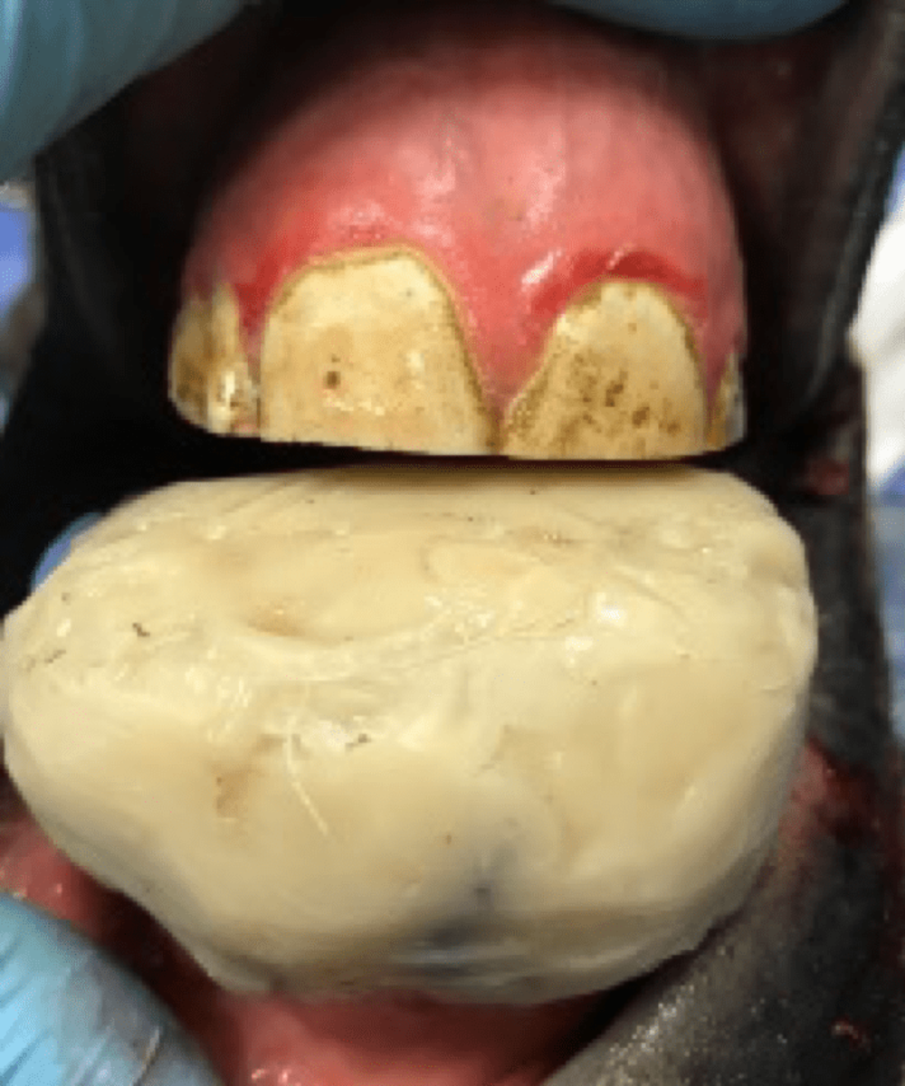
This image shows the “hooks” that can appear when the top and bottom jaws don't align properly and part of a tooth is not worn down as normal. Photo courtesy Dr. Brad Tanner

This can cause significant problems and pain for horses, as these large hooks cause collateral damage to the soft tissues, gums, and cheeks. This is compounded when a bit is applied to the horse's mouth and a rider asks the horse to put the head into a frame/collection, causing increased contact of these teeth overgrowths and the soft tissues surrounding them. In Thoroughbred racehorses the effects are less dramatic than in dressage and other disciplines. This is due to the fact that racehorses run largely on looser reins with an extended head and neck rather than in flexion of the poll.

Can it be corrected?

While this is a permanent deformity in adult horses, it can be corrected in young growing foals. Correction should take place between four to six months of age, ideally. As the maxilla (top jaw) is growing ahead of the mandible (lower jaw) at a higher rate, an orthodontic appliance can be applied with orthodontic wires to halt the growth of the maxilla allowing the mandible to catch up. Additionally, an inclined plane is applied in the form of an aluminum plate allowing the lower teeth to engage with it during each chewing motion.

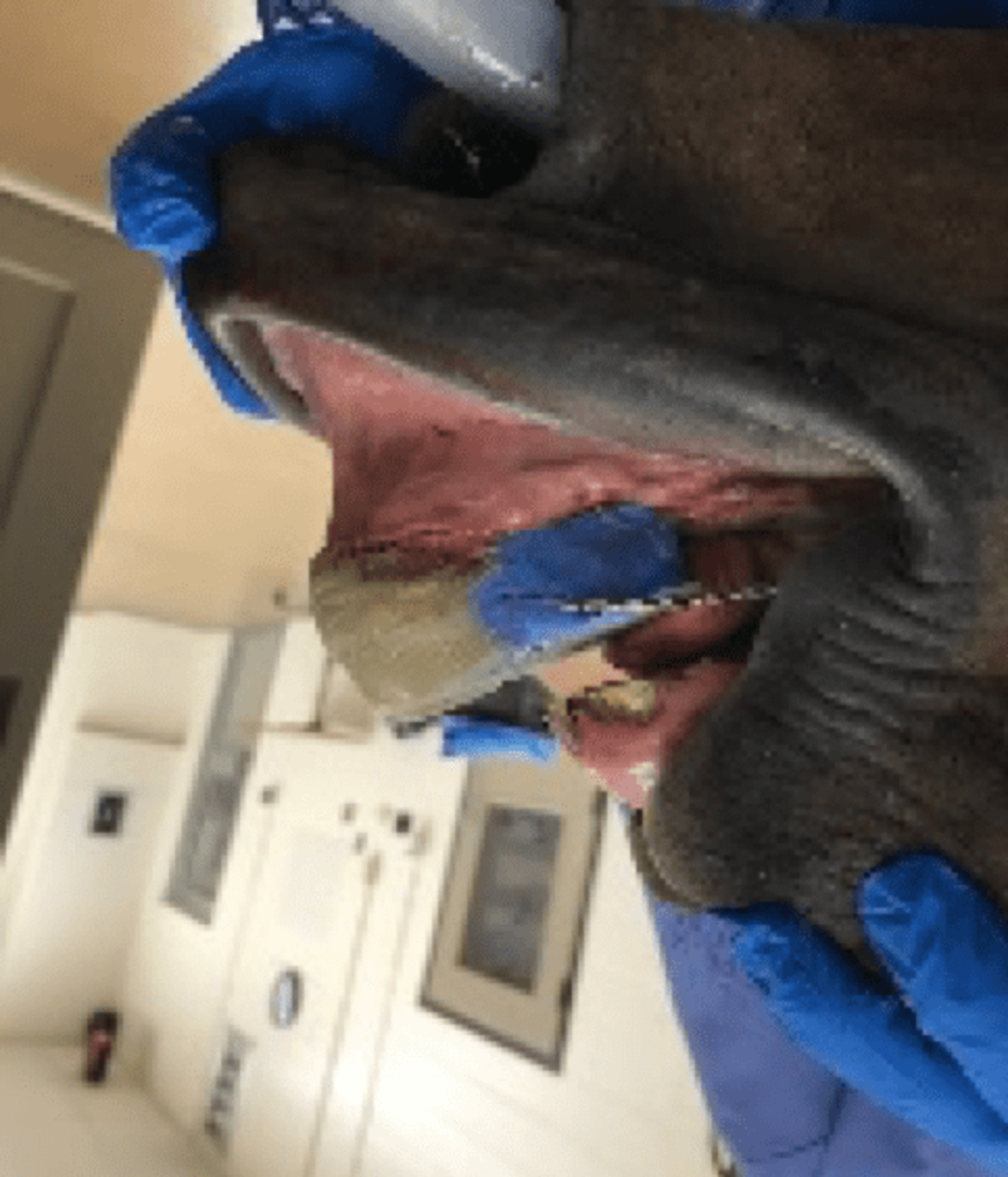
Corrective devices can fix parrot mouth if it is caught early in a horse's life, before skeletal development is complete. Photo courtesy Dr. Brad Tanner

Corrective devices can fix parrot mouth if it is caught early in a horse's life, before skeletal development is complete. Photo courtesy Dr. Brad Tanner

Once the skeletal development of the horse is complete, there is no way to effectively correct the overbite/parrot mouth malocclusion.

How do you manage this problem long term?

Long term management of these malocclusions should be focused on effectively floating and reducing the large overgrowths. These are the overgrowths that form because some teeth are not in occlusion and chewing along one another. Normally, the teeth will grind and wear out at the same rate but when one tooth is not in contact it is allowed to continue to grow through the life of the horse at a rate of three to four millimeters per year. Left unchecked, this can wreak havoc on the horse's mouth and be a painful situation.

Long term management often will require routine floating examinations at six-month intervals on horses that have these types of malocclusions. So long as these are managed effectively the horse will be able to have normal dentition and perform at a normal level. Horses will not run faster or slower because of the parrot mouth. So long as they are managed effectively it should have no influence on performance.

The heritability of a parrot mouth has been called into question for some time. It's often viewed as a heritable trait that can be passed along from generation to generation and is therefore undesirable in breeding stock. Unfortunately, while much research has been done in this area, the heritability of overbites/parrot mouth is a very complex genetic trait. Recent studies have shown there are complex patterns of genetic alleles that are involved in the expression of the genes that lead to this defect. It is difficult to pinpoint exactly which foals could develop this trait and which sires are more or less prone to passing it along because of the complexity of alleles that are necessary to give this result and deformity.

In summary, parrot mouths are not really a big deal for a lot of horses. They have managed to survive for millennia this way and will continue to eat and graze. Proper management of a mature horse with parrot mouth will lead to decreased dental problems, decreased pain, and better performance in a show ring or racetrack. Recognition at an early age does allow for surgical correction and normal dentition for the rest of the horse's life.

Dr. Brad Tanner received his DVM from Auburn University in 2005 and completed an internship at Rood and Riddle thereafter. He is a shareholder in the clinic, where he focuses on advanced dentistry, primary care and reproduction. Tanner became board certified in equine dentistry in 2019.

The post Ask Your Veterinarian Presented By Kentucky Performance Products: What To Do About ‘Parrot Mouth’ appeared first on Horse Racing News | Paulick Report.

Source of original post

Ask Your Veterinarian Presented By Kentucky Performance Products: What’s Bone Bruising?

Veterinarians at Rood and Riddle Equine Hospital answer your questions about sales and healthcare of Thoroughbred auction yearlings, weanlings, 2-year-olds and breeding stock.

QUESTION: What does it really mean when a veterinarian talks about a racehorse having “bone bruising”?

DR. A.J. RUGGLES: If you been around racehorses you likely have heard the term “bone bruising.” Despite its common use the term is really not entirely accurate in most cases. What your veterinarian is likely referring to is Non-Adaptive Stress Remodeling (NASR). You can see why the term bone bruising is more commonly used.  While a true contusion (bruise) of the bone–manifested by lameness and characteristic findings of edema in the bone on magnetic resonance imaging–occurs, it is much less common than NASR.

To understand NASR and its causes, an understanding of bone anatomy and physiology is necessary. Most people think of the skeleton as an inert frame that muscle, tendon and ligaments attach to allow movement or as protection for vital organs. While the skeleton performs these functions, it also is a very dynamic system than is undergoing a constant process of removal and replacement as the horse grows in size and is being trained.

Dr. Alan Ruggles

It is easy to realize the skeleton of a foal is different than the skeleton of a 3-year-old. Not only has the horse grown in stature, but the structure of the bone itself is altered to fit its athletic activity. For example, the front of the cannon bones in the front legs of a trained 3-year-old will be thicker and denser compared to a 3-year-old that has never trained and only exercised at pasture. Likewise, an older broodmare who has been out of training and has had many foals may have a relatively weaker skeleton compared to the actively trained racehorse due to the absence of training and the depletion of calcium from her skeleton due to multiple lactation cycles.

When an athlete trains, whether it is a person or horse, receptors between the cells within the bone recognize the changes in load in the bone and send a signal for the bone to change its geometry and replace damaged bone to fit this new activity. This normal process is called stress remodeling. During this process original bone is removed and new bone is produced to replace it. Imagine a long bone like the cannon bone as a cable of a suspension bridge and within the cable are multiple smaller wires cables.

When bone changes its shapes or repairs injured bone each of these original smaller wires (primary osteons) are removed and then replaced with new bone (secondary osteons). During this process the removal phase occurs at a rate 50 times faster than the replacement rate. The rate of remodeling is influenced by the stimulus of training and when it occurs successfully, the process is necessary and positive. Another response of bone is to make itself larger quickly to resist mechanical loads by putting down relatively weaker (woven not cortical bone) on the surface of a bone. This is what causes the bump in bucked shins.

Most of the adaptive process of the horse skeleton via stress remodeling occurs without incident but sometimes the process gets overwhelmed and manifests as lameness. If there is a failure of the normal stress remodeling process, there can be an accumulation of damaged bone which can cause lameness, micro fissures and fractures.

An image captured from a bone scan shows an area of concern

Sometimes the lameness is obvious and easily detected, such as a bucked shin of the front cannon bone. Sometimes the lameness is obvious but not easily detected on a physical exam as with humeral, tibial or ilial stress fractures. Most commonly, at least in our practice, the horse has clinical signs of poor performance: perhaps a “crabby gait” or not changing leads. The horse is often lame in more than one limb, which makes detection of the problem more difficult.

A careful lameness examination with diagnostic nerve blocks is recommended to ferret out the cause of the lameness. The nerve blocks help us localize the source of lameness and help us direct our diagnostic imaging such as radiographs, ultrasound, nuclear scintigraphy, MRI or computed tomography.

Nuclear scintigraphy (commonly known as bone scan) is very helpful in detecting stress remodeling since it is best suited to detect excess bone metabolic activity which occurs during stress remodeling and stress fractures.

Radiographs and computed tomography may reveal increased density of the bone with associated bone resorption especially in the condyles of the distal cannon bone. There also may be changes in the bone contour and the development of fractures. Magnetic resonance imaging with high field magnets (MRI) is helpful to determining the health of the cartilage as well as bone and associated soft tissue structures. Ultrasound is not generally helpful in diagnosis or management. Newer technologies such as standing computed tomography and PET scans give detailed 3D images of bone and show promise but are not yet available for widespread use and are still undergoing clinical validation in the management of stress remodeling.

An MRI shows an area of bone bruising

If your horse is diagnosed with bone bruising, it likely has a form of NASR. That is the bad news. The good news is most cases do not develop clinical fractures and therefore are not treated surgically and responds to rest. Horses that develop fractures such as dorsal cortical fracture of the cannon bone or condylar fractures of the cannon often are treated surgically for best outcomes. Other hairline fractures of the humerus and tibia are treated with rest alone. The majority of cases in racehorse affect the bottom of the cannon bone or the third carpal bone and are treated with rest.

An image from a radiograph shows bone bruising

Typically, they are given 60 to 90 days off and pasture activity is recommended. The purpose of the period of rest is to allow the skeleton to catch up with the signals that have been sent by training so the stress remodeling process can finish to better allow the bone to withstand the rigor of training and racing. Remember, during stress remodeling bone resorption is 50 times faster than production.

Timing on when to turn the horse out obviously depends on the degree of lameness. If the horse shows any potential for a fracture being present, negative follow-up radiographs are needed before turnout etc. All these decisions are unique for each horse and should and be made in concert with your veterinarian. Treatments such aspirin and isoxsuprine and some over the counter supplements may help during the process by improving blood flow to the bone. Drugs which inhibit the natural bone remodeling process such as bisphosphonates, in my opinion, should not be used in cases of NASR and should only be used as labeled.

The vast majority (80% plus) respond to a period of rest with turnout. This is a tried and true method of trainers who have for decades given horses time off usually in the winter. These methods still work today of course. Proper diagnosis of NASR is important in my opinion to make sure you know what you are treating and to make sure no other condition exists that might require a different intervention.

Next time you hear that a horse has bone bruising, remember it is likely a form of NASR and hopefully you will have a better understand of the natural process of bone turnover and how it is related to this syndrome.

Dr. Ruggles specializes in orthopedic surgery and lameness. In addition to his experience as a practicing veterinarian, he served as a faculty member at New Bolton Center and at Ohio State University before joining Rood and Riddle in 1999. He is a partner in the hospital and is part of the AAEP “On Call” media program.

The post Ask Your Veterinarian Presented By Kentucky Performance Products: What’s Bone Bruising? appeared first on Horse Racing News | Paulick Report.

Source of original post

Verified by MonsterInsights